Clinical example No. 41. Diagnosis: fixed postlaminectomic kyphotic deformation of the cervical spine (laminectomy, tumor removal in 1988), combined stenosis of the cervical spine at the level of C4, C6, C6 of the vertebrae – the Federal Center for Neurosurgery, Novosibirsk
The main page
Clinical example No. 41. Diagnosis: fixed postlaminectomy kyphotic deformation of the cervical spine (laminectomy, tumor removal in 1988), combined stenosis of the cervical spine at the level of C4, C6, C6 vertebrae
Patient N., 58 years old, entered the Department of Spinal neurosurgery of the Fats of Novosibirsk with complaints on weakness in the limbs, impaired movements in the fingers of the left hand, restriction of movements in the cervical spine, forced position of the head and cervical spine.
From the anamnesis it is known that in 1988, surgical treatment was performed-laminectomy C4, C5, C6, removal of neurinoma at the level of C4-C6. Since that time, he noted the progressive kyphotic deformation of the cervical spine. In 2014, for the first time, weakness appeared in the left hand with the gradual progression of the degree of weakness with the appearance of weakness in the remaining limbs
Orthopedo-neurological status: tetraparesis, upper peripheral, lower in central type, up to 4 points, rude in the left hand up to 3 points with the formation of combined contracture in the left wrist joint; Reflexes on the upper extremities are reduced, symmetrically, in the lower ones are lively with the expansion of reflexogenic zones with pathological stop signs; Increased muscle tone of the lower extremities (Joa 13, Frankel D, Ashvord 2).
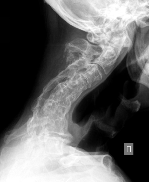
Upon admission, the cervical spine is performed, Figure 1.

Figure 1. The lateral radiograph of the cervical spine of the patient N.
Kifotic deformation of the cervical spine is visible with the formation of a bone block on the lower cervical segments.
To clarify the morphological changes in the bone structures, the vascular-naval structures also made an MRI of the cervical spine, Figure 2 and MSCT of the cervical spine, Figure 3.

Figure 2. MRI of the cervical spine of the patient N.
According to the results of the MRI, the continued tumor growth is not determined. On the sagittal cut, the zone of myeloichemia of the spinal cord is visible with the formation of a syringomyelic cyst at the level of C4 the vertebra, the postoperative cyst, and gross deformation of the spinal cord is visible in axial images.